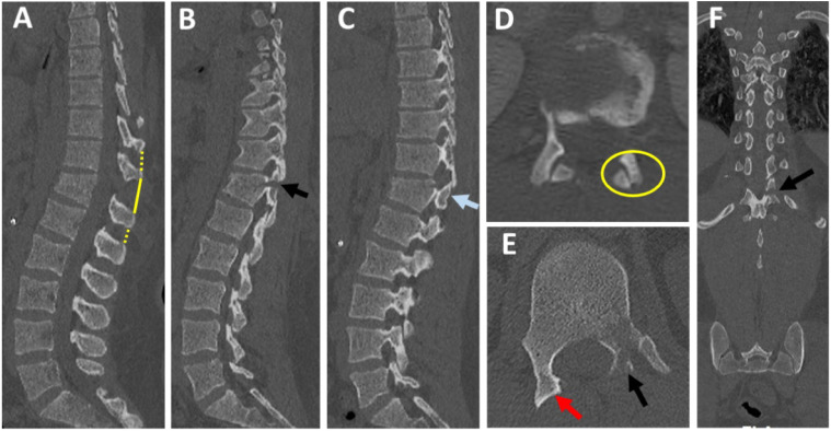
Figure 2.
CT checklist for diagnosis of PLC injury. (A) Midsagittal CT shows should be checked for interspinous widening (yellow solid and interrupted lines, as well as vertebral translation and spinous process fracture; (B-C) parasagittal CT, shows left laminar and pedicle fracture (B, black arrow) and right subluxed facet (C, blue arrow); (D-E) Axial CT image shows right subluxed facet (D, yellow ring) and naked facet sign (E, red arrow) and left laminar, and pedicle fracture (E, black arrow); (F) coronal reconstruction image shows the horizontal orientation of the laminar fracture (black arrow).

