Fig. 2.
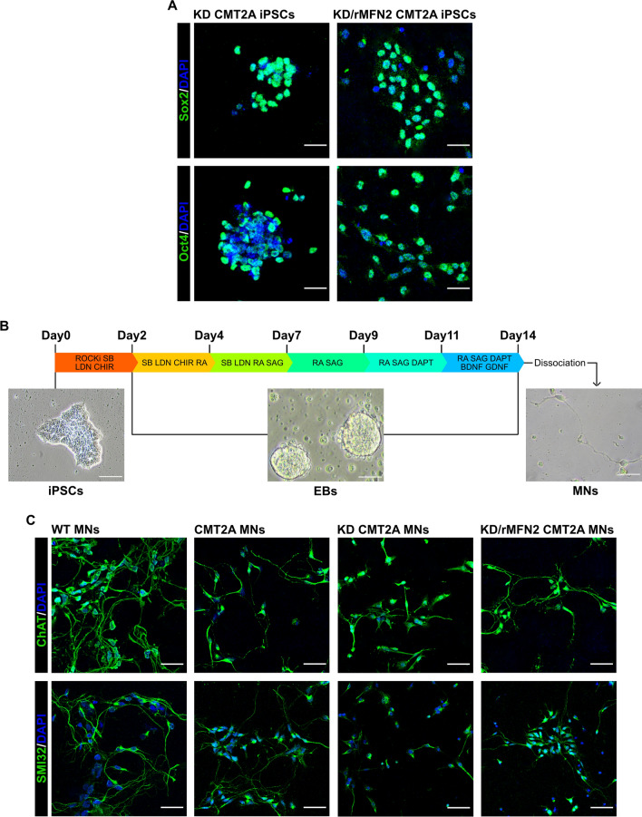
Combined RNAi/gene therapy preserved iPSC and MN phenotypes. A Immunocytochemistry of KD-CMT2A and KD/rMFN2-CMT2A iPSCs for pluripotency transcription factors SOX2 (green) and OCT4 (green). Nuclei were labeled with DAPI (blue). Images were acquired using a Leica TCS SP5 confocal microscope at 20 × magnification. Scale bar: 25 μm. B Experimental outline for MN differentiation of WT, CMT2A, KD-CMT2A, and KD/rMFN2-CMT2A iPSCs and bright-field representative images at three different differentiation time points (iPSC, EB, and MN). Scale bars: 100 μm for iPSCs, 50 μm for both EBs and MNs. C Immunocytochemistry of WT, CMT2A, KD-CMT2A, and KD/rMFN2-CMT2A MNs after 2 weeks of differentiation for MN markers SMI32 (green) and ChAT (green). Images were acquired using a Leica TCS SP5 confocal microscope at 20 × magnification. Nuclei were labeled with DAPI (blue). Scale bar: 50 µm

