Fig. 5.
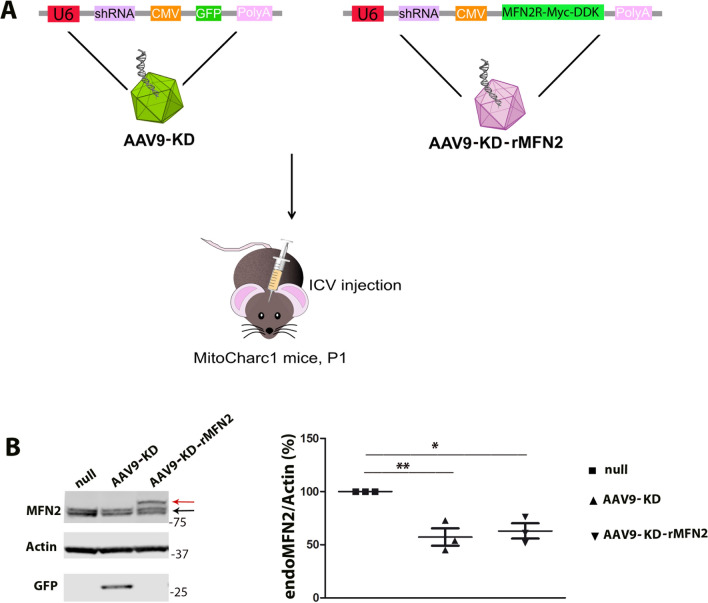
AAV9-KD and AAV9-KD-rMFN2 efficiently transduced tissues in CMT2A mice. A In vivo experimental design. Newborn (P1) CMT2A were injected intracerebroventricularly (ICV) with AAV9-KD or AAV9-KD-rMFN2. B Representative Western blot of endogenous MFN2 protein (endoMFN2, black arrow) and exogenous MFN2 (Myc-DDK MFN2, shift of higher molecular weight, red arrow) after AAV9-KD, AAV9-KD-rMFN2 or AAV9::null treatment in the brains of MitoCharc1 mice. AAV9-KD transduction in the brains was confirmed by the detection of GFP tag. Densitometric quantification (n = 3). Error bars indicate SEM of endoMFN2/actin expression. **P < 0.01,*P < 0.05, Student’s t-test

