
Figure 2. SIIN-Q11 i.n. immunization results in situ priming and proliferation of naive CD8+ T cells in the lung before draining MLN.
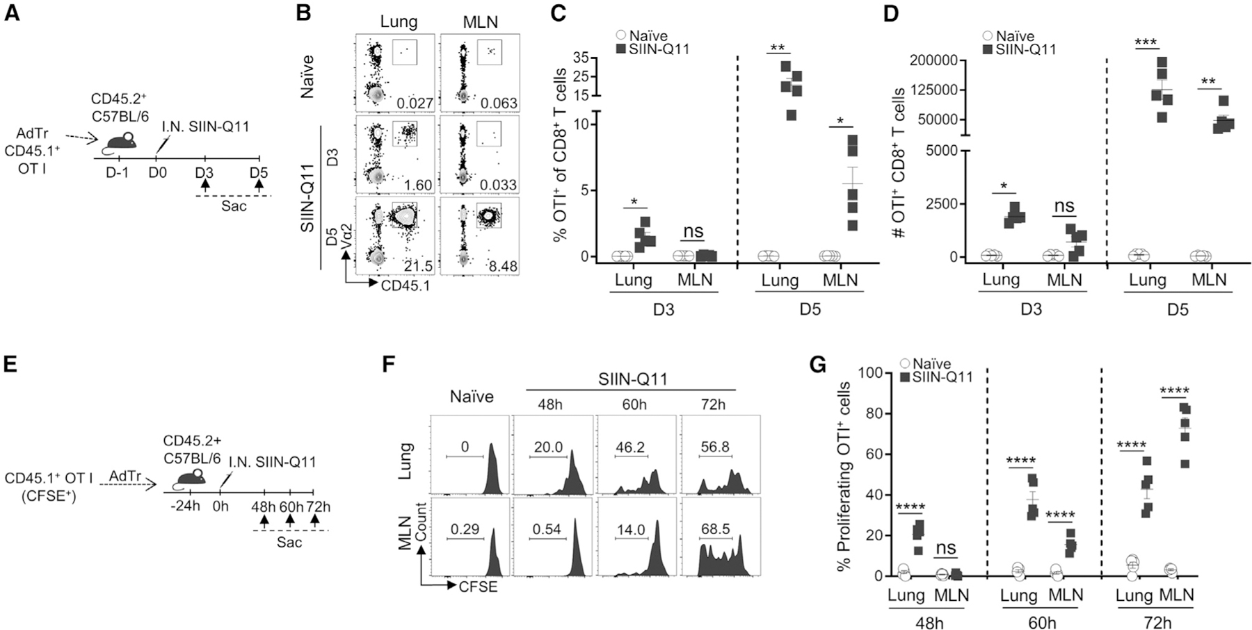
(A) Experimental design for (B)–(D). OT-1 T cells were purified from OT-1 TCR-Tg CD45.1+ mice and adoptively transferred to CD45.2+ C57BL/6 mice (50,000/mouse) at 24 h before immunization. Mice were sacrificed on day 3 (D3) or day 5 (D5) post immunization. OT-1 T cells were identified as CD45.1+Va2+CD8+ T cells. OT-1 cell accumulation in the lung vs. MLN was analyzed.
(B) Representative flow cytometry plots identifying OT-1 cells in lung (left) and MLN (right).
(C and D) (C) Percentage of OT-1 T cells of CD8+ T cells, and total number (D) in lung and draining MLN on day 3 vs. day 5 after immunization.
(E) Experimental design for (F) and (G). CD45.1+ OT-1 cells were labeled with CFSE prior to adoptive transfer to CD45.2+ C57BL/6 mice (50,000/mouse) at 24 h before immunization. Mice were sacrificed and lungs and MLNs were harvested at the indicated time points after immunization. Proliferation of adoptively transferred OT-1 cells were analyzed by flow cytometry for CFSE dilution.
(F) Representative flow cytometry plots showing CFSE dilution of OT-1 T cells in the lung (top) and MLN (bottom) at different time points.
(G) Percentage CFSE-diluted cells of OT-1 cells at 48, 60, and 72 h after immunization. Each dot represents one mouse. Data shown are means ± SEM from two independent experiments (C, D, G). ****p < 0.0001; ***p < 0.001; **p < 0.01; *p < 0.05; ns, not significant by two-way ANOVA (C, D, G).