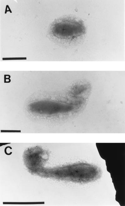
FIG. 6.
Synchronized VP-6 cells labeled with cationic ferritin. Cells were removed from a synchronous culture, treated with a 1.0-mg/ml solution of cationic ferritin, washed, and layered onto collodion-coated copper electron micrograph grids. Cells shown in these photomicrographs were removed from the synchronous culture at 0 min (A), 180 min (B), and 360 min (C). Notice that VP-6 produced capsular EPS at each of the time points. Bar = 1 μm.

