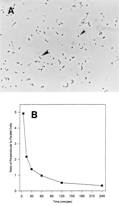
FIG. 8.
Perpendicular versus parallel adhesion. (A) Phase-contrast image of VP-6 cells attached to a glass coverslip. Perpendicularly attached cells appear spherical (small arrowhead) and can be easily differentiated from those lying parallel to the surface (large arrowhead). Magnification, ×925. (B) Ratio of perpendicular to parallel cells relative to time of adhesion. The ratio of cells exhibiting perpendicular adhesion to those exhibiting parallel adhesion decreases with time.

