. 2020 Apr 12;147(8):dev184655. doi: 10.1242/dev.184655

Fig. 1.

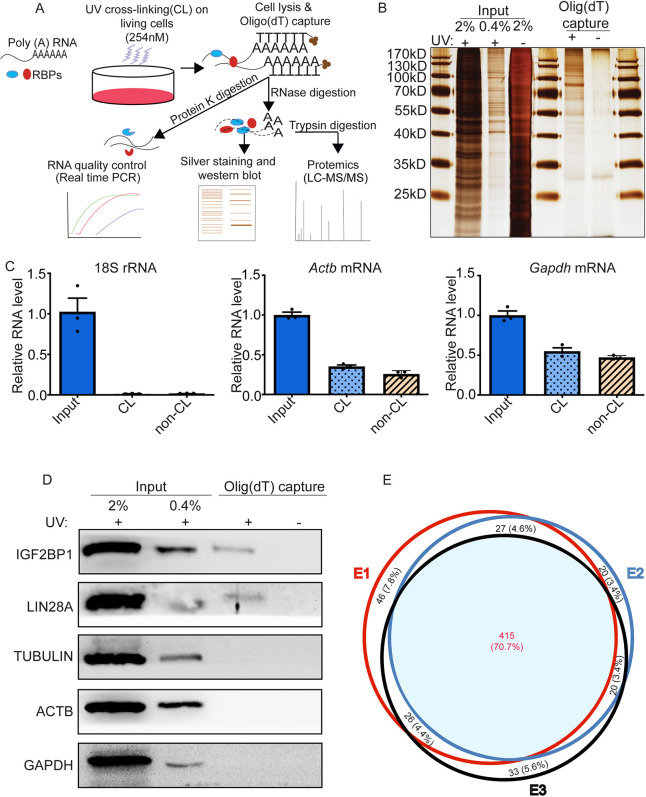
Fig. 1. mRBPome profiling of mouse undifferentiated spermatogonia. (A) Schematic of the experimental approach. mRNA-protein complexes in primary cultures of undifferentiated spermatogonia were UV-crosslinked and captured using oligo(dT) magnetic beads. (B) Representative image of a silver-stained SDS-PAGE gel confirming enrichment of protein bands in UV-crosslinked (CL; +) compared with non-UV-crosslinked control (non-CL; −) input and oligo(dT)-captured samples. (C) Relative levels of 18S rRNA, Actb and Gapdh mRNAs measured by quantitative real-time RT-PCR analysis. Data are mean±s.e.m. for n=3 different cultures. (D) Representative image of western blot analysis confirming enrichment of the known RBPs IGF2BP1 and LIN28A in UV-crosslinked samples subjected to oligo(dT)-capture (+). (E) Venn diagram analysis of RBPs identified by mass spectrometry analysis; data are from three independent primary cultures.

mRBPome profiling of mouse undifferentiated spermatogonia. (A) Schematic of the experimental approach. mRNA-protein complexes in primary cultures of undifferentiated spermatogonia were UV-crosslinked and captured using oligo(dT) magnetic beads. (B) Representative image of a silver-stained SDS-PAGE gel confirming enrichment of protein bands in UV-crosslinked (CL; +) compared with non-UV-crosslinked control (non-CL; −) input and oligo(dT)-captured samples. (C) Relative levels of 18S rRNA, Actb and Gapdh mRNAs measured by quantitative real-time RT-PCR analysis. Data are mean±s.e.m. for n=3 different cultures. (D) Representative image of western blot analysis confirming enrichment of the known RBPs IGF2BP1 and LIN28A in UV-crosslinked samples subjected to oligo(dT)-capture (+). (E) Venn diagram analysis of RBPs identified by mass spectrometry analysis; data are from three independent primary cultures.