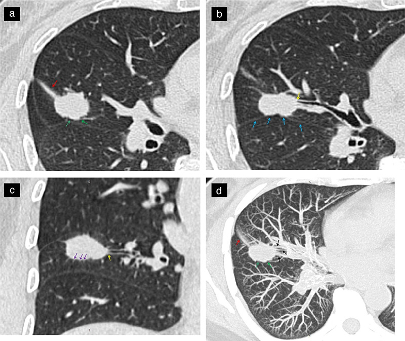
Supplementary Figure 1.

Show a same patient, a 53-year-old female patient with lung adenocarcinoma and positive STAS status. The axial non-contrast computed tomography shows a solid nodule in the right middle lobe of the lung, CTR ≥50%, with distal ribbon sign (a, red arrow), lobulation sigh (a, green arrow), interlobar pleura indentation sigh (b, blue arrow), bronchial change (b and c, yellow arrow), spiculation sigh (c, purple arrow). The maximal intensity projection (d) shows a vascular convergence sign (black arrow), lobulation sigh (green arrow) and distal ribbon sigh (red arrow). STAS, spread through air spaces; CTR, consolidation-to-tumor ratio.
