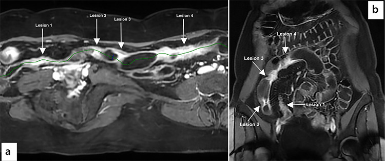
Figure 2.

Magnetic resonance enterography (a) reveals small bowel alteration affecting the last 40 cm of the ileum, characterized by the presence of four thick-walled and reduced-caliber tracts, which are represented in the reconstructions obtained through vessel analysis. In this reconstruction, it is possible to accurately assess the overall extent of the disease and each lesion. Lesion 1: length of 70 mm, lesion 2: 10 mm, lesion 3: 14 mm, lesion 4: 100 mm. Coronal T1 image performed after the intravenous injection of gadolinium contrast medium (b) does not allow the small bowel to be visualized in its longitudinal extension because of the long section of intestine affected by the pathology; however, it was possible to measure the length of each pathological loop in the various planes.
