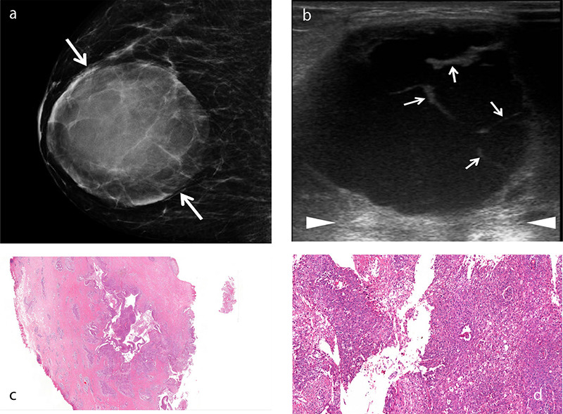
Figure 1.

An example of squamous cell metaplastic breast carcinoma with extensive central necrosis. A 38-year-old female patient presented with a rapidly growing palpable mass in the right breast. Mammogram (a) reveals a large, well-defined round density mass measuring 8 × 7.5 cm with circumscribed margins (arrows). Gray-scale ultrasound image (b) shows a complex cystic mass with thick septa (b, arrows) and posterior acoustic enhancement (arrowheads). Histopathology of the cystic lesion (c) reveals atypical squamous cells lining the cavity and infiltrating the adjacent stroma [hematoxylin and eosin (H&E), ×2]. Photomicrograph (d) shows that the neoplastic cells lining the irregular spaces are atypical squamous cells with abundant eosinophilic cytoplasm and large, vesicular nuclei (H&E, ×20).
