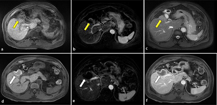
Figure 2.

Comparison of pre- and post-treatment imaging in a 70-year-old male with a history of hepatocellular carcinoma (HCC) in the context of chronic hepatitis B after microwave ablation of residual disease in segment 5 sixteen months after trans-arterial chemoembolization. Pre-treatment magnetic resonance imaging (MRI) T1-weighted sequences with (a) non-contrast, (b) arterial, and (c) portal venous phases show a residual 3.5 cm nodular enhancing segment-5 lesion with peripheral washout at the anterosuperior and left lateral borders of the treatment cavity consistent with Liver Imaging Reporting and Data System Treatment Response viable (yellow arrow). Post-treatment MRI T1-weighted sequences with (d) non-contrast, (e) arterial, and (f) portal venous phases demonstrate a new ablation cavity with no evidence of arterial phase hyperenhancement or washout consistent with LIRADS-TR non-viable (white arrow).
