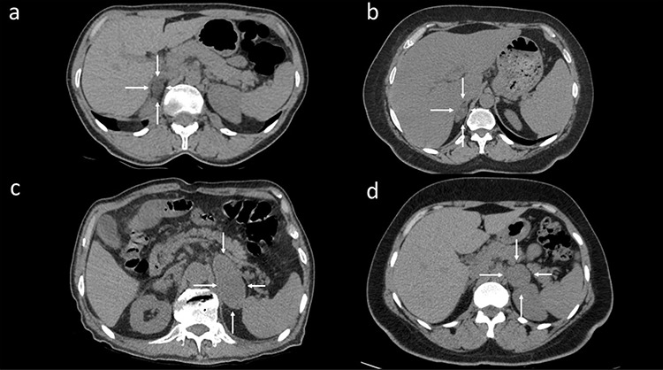
Figure 2.

(a) Lipid-rich adenoma in a 49-year-old woman. A well-circumscribed and low-density lesion was observed in the right adrenal gland in axial computed tomography (CT) (white arrows). (b) A 37-year-old woman with lipid-poor adenoma diagnosed histopathologically. Axial unenhanced CT showed a lipid-poor right adrenal mass (white arrows). The mass with a density of 22–56 Hounsfield unit (HU) had a heterogeneous internal structure. (c) Adrenal metastases were diagnosed through surgery in a 56-year-old man with lung cancer. Axial unenhanced CT showed a large, well-defined, homogenous left adrenal mass with a density of 54 HU (white arrows). (d) Surgically resected pheochromocytoma in a 32-year-old man. Axial unenhanced CT showed a well-defined, homogenous, spherical left adrenal mass with a density of 46 HU (white arrows).
