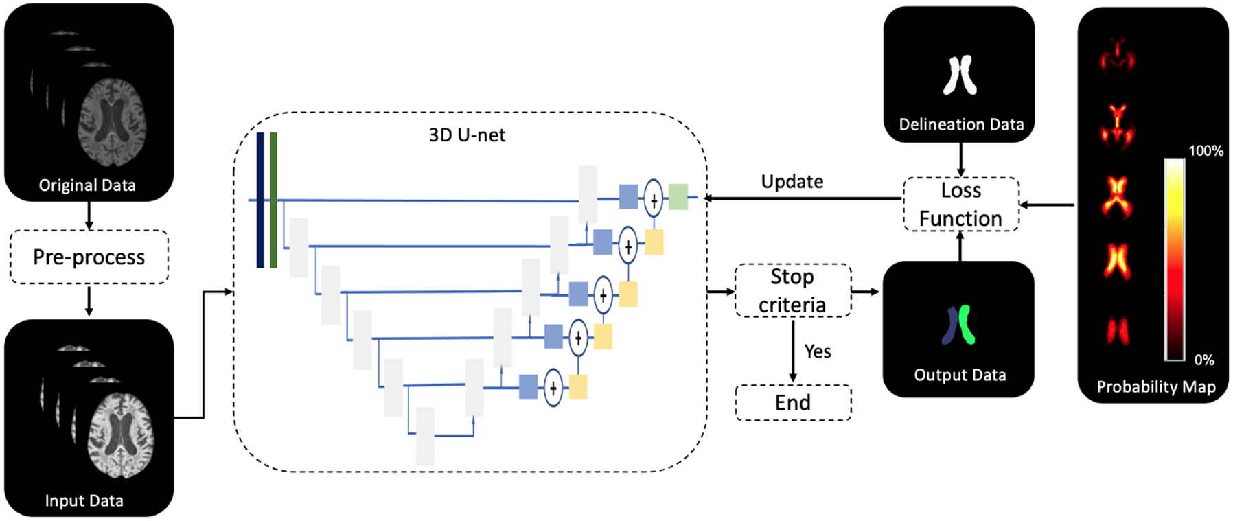
Figure 1.

Flowchart for MRI ventricle segmentation. Segmentation of the MRIs is achieved with a modified 3D U-Net. The input data to the 3D U-Net is pre-processed from the original data. The loss function used to train the network consists of the DSC loss of all labels between ground truth delineation data and network output data, as well as the binary cross-entropy loss between the probability map and network output data.
