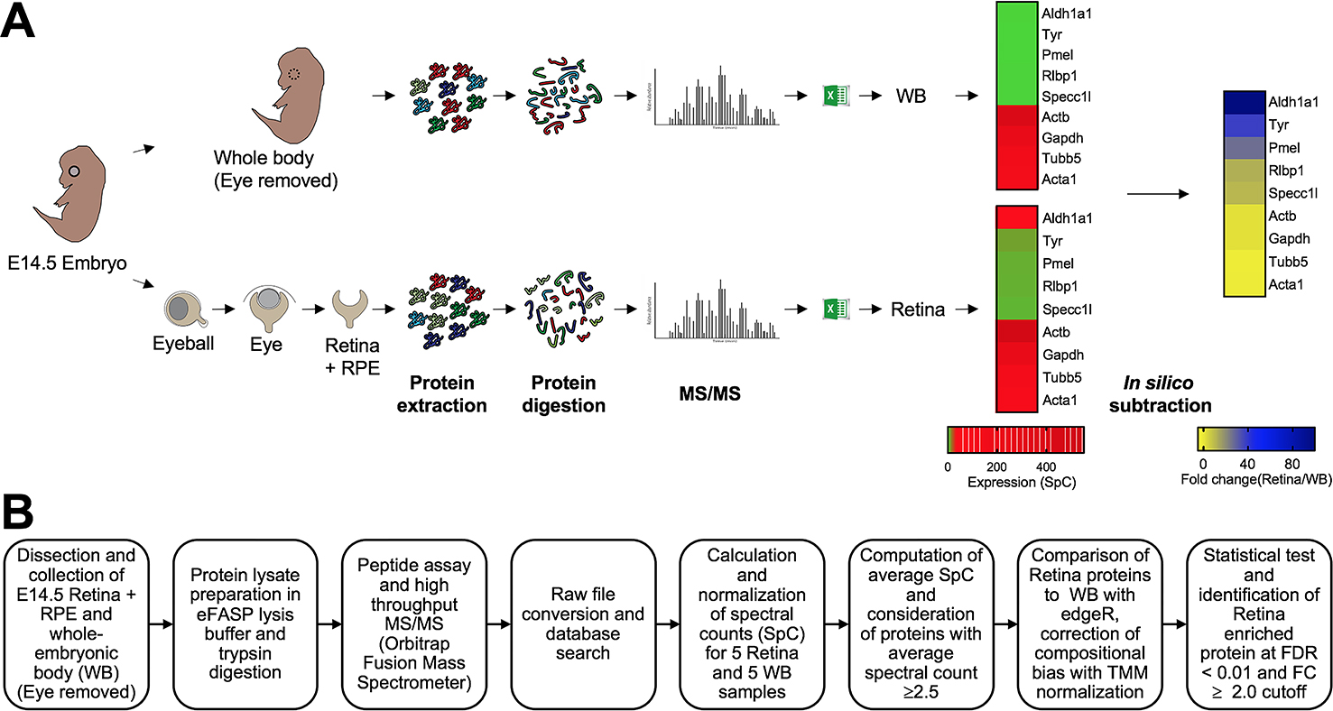
Fig. 1. Workflow of the experimental strategy to generate MS/MS protein profile of the mouse embryonic retina and retinal pigment epithelium combined tissue.

(A) Mouse eyes at embryonic day (E)14.5 were isolated, and the retina and retinal pigment epithelium combined tissue (termed retina) was micro-dissected. The whole body (WB) with eye tissue removed was processed similarly and used as reference for differential protein expression analysis. Retina and WB samples (n = 5 for each sample type, 55 μg protein per sample) were subjected to high-throughput tandem mass spectrometry (MS/MS). (B) The workflow for differential protein expression analysis is outlined. The edgeR pipeline was used to determine differential protein expression using normalized spectral counts. Proteins passing stringency criteria of ≥2.5 average spectral counts, ≥2.0 fold-change (in retina, compared to WB), False Discovery Rate <0.01 were considered to have enriched expression in the retina.
