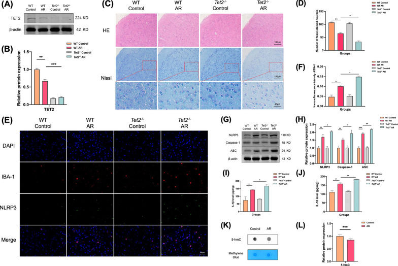
Fig. 4.
TET2 depletion aggravates nerve injury in ACC of AR mice. A Expression levels of TET2 in the ACC were determined using western blot analysis. B Quantitative analysis of band intensity. C Representative HE-stained and Nissl-stained images of the mice ACC. D Number of Nissl-stained neurons. E Representative immunofluorescence images of colocalization of NLRP3 in microglia in ACC. F Immunofluorescence of intensity of IBA-1. G Expression levels of NLRP3, Caspase-1, and ASC in the ACC were determined using western blot analysis. H Quantitative analysis of band intensity. I, J Levels of IL-1β and IL-18 were determined by ELISA in the ACC. K Levels of 5-hmC determined by dot blot in the ACC. L Quantitative analysis of 5-hmC levels in the ACC. Data are presented as mean ± SEM. (*P < 0.05, **P < 0.01, ***P < 0.001)

