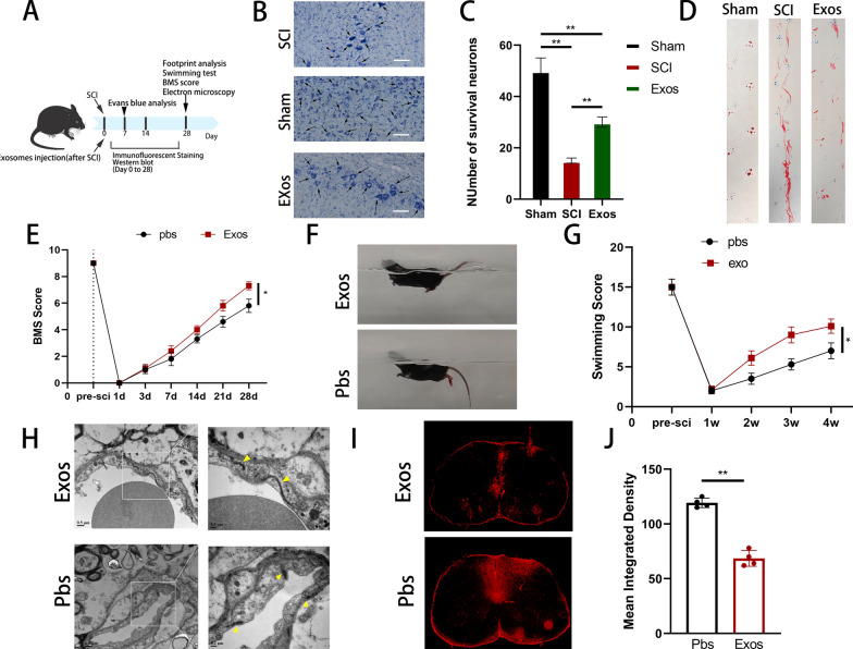
Fig. 4.
Pericyte-derived exosomes promote the recovery of motor function and protect BSCB in mice after spinal cord injury. A Schematic diagram of the experiment after spinal cord injury in mice. B, C Nissl staining showed and quantified the number of mouse motor neurons in each group. D Representative footprints of walking 28 days after spinal cord injury and quantifying the results of footprint analysis in individual mice. Blue: Forefoot; Red: Backfoot. E Functional classification of mice in each group using BMS scores from pre-injury to 28 days post-injury. F, G Functional grading at 28 days post-injury using the Louisville Swim scale and representative images. H The morphology of vascular tight junctions on the 28th day after spinal cord injury in different groups of mice was observed by transmission electron microscope. I, J Representative fluorescent images and quantification of spinal cord cross-sections in the injured area after EB injection at 7 days post-injury

