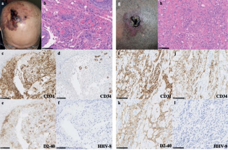
Fig. 1.
Clinical and histological findings. (a) Mass and purpura on the right crown region of the scalp. (b) Haematoxylin and eosin (H&E) staining of a biopsy specimen of the mass revealed atypical spindle-shaped cells forming irregular vessels in the dermis. (c–f) Tumour cells were positive for CD31 and D2-40 and negative for CD34 and human herpesvirus-8 (HHV-8). (g) Plaque on the left frontal region of the scalp. (h) H&E staining of a biopsy specimen showed atypical cells forming dilated irregular vascular structures in the dermis. (i–l) Tumour cells were positive for CD31, CD34 and D2-40 and negative for HHV-8. Bars: 100 μm.

