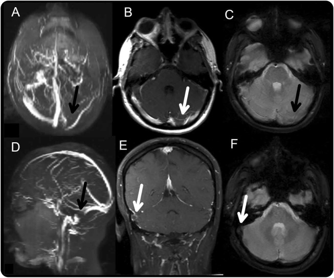
Figure. Cerebral venous sinus thrombosis.
(A) Coronal magnetic resonance venography reconstruction of the patient's cerebral veins, obtained on the fourth day of admission. Absence of flow in the left transverse sinus (black arrow) is shown. (B) Contrast-enhanced T1 MRI demonstrates lack of flow in the left transverse sinus (white arrow). (C) Gradient echo image shows relative decreased susceptibility in the transverse sinus consistent with thrombosis but no hemorrhage. (D) Sagittal magnetic resonance venography reconstruction of the patient's cerebral veins shows absence of flow in the right sigmoid sinus (black arrow). (E) Contrast-enhanced T1 MRI demonstrates absence of contrast filling in the right sigmoid sinus (white arrow). (F) Gradient echo image shows decreased susceptibility in the right sigmoid sinus (white arrow).

