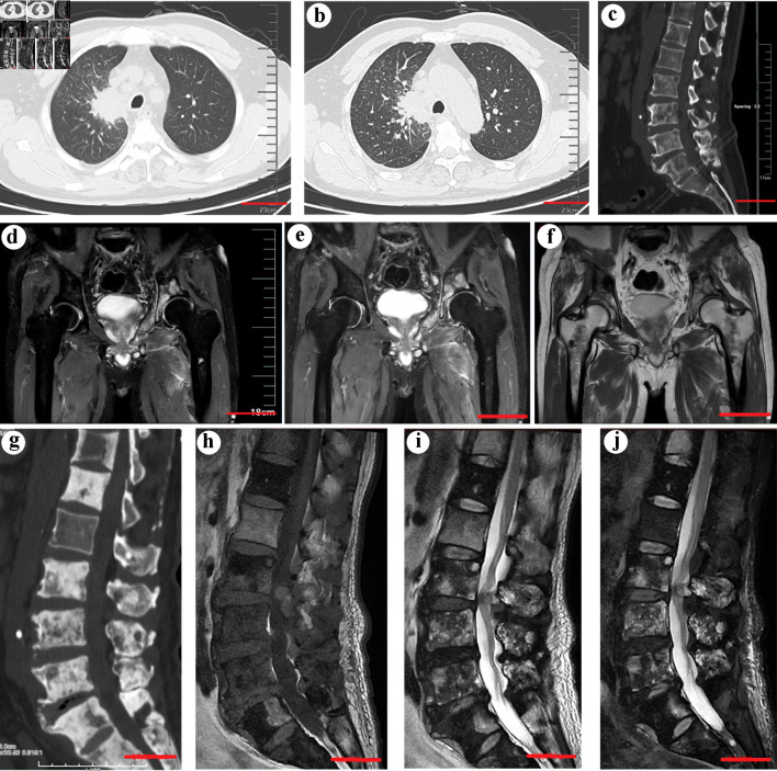
Figure 1.
(a) Computed tomography (CT) scan showing a neoplasm in the right upper lobe (scale bar = 5 cm). (b) An enhanced CT scan confirmed the existence of a neoplasm (scale bar = 5 cm). (c) CT scans of the spine before treatment with sintilimab, bevacizumab, and chemotherapy showed multiple osteolytic lesions in the thoracic and lumbar vertebrae (scale bar = 5 cm). (d, e, f) An MRI study showed new pelvic and bilateral femoral metastases (scale bar = 5 cm). (g) CT scans of the spine showed marked osteoblastic activity and osteosclerosis in the vertebra with metastases after the treatment (scale bar = 5 cm). (h, i, j) The corresponding MRI scans of the spine after treatment. (h) T1W sagittal spin-echo sequence of the spine showed hypointense lesions in multiple thoracic and lumbar vertebrae. The T2W sagittal spin-echo sequence (i) and T2W sagittal spin-echo sequence with fat suppression (j) showed both hyperintense and hypointense lesions in multiple thoracic and lumbar vertebrae, which indicates osteoblastic bone lesions (scale bar = 5 cm).

