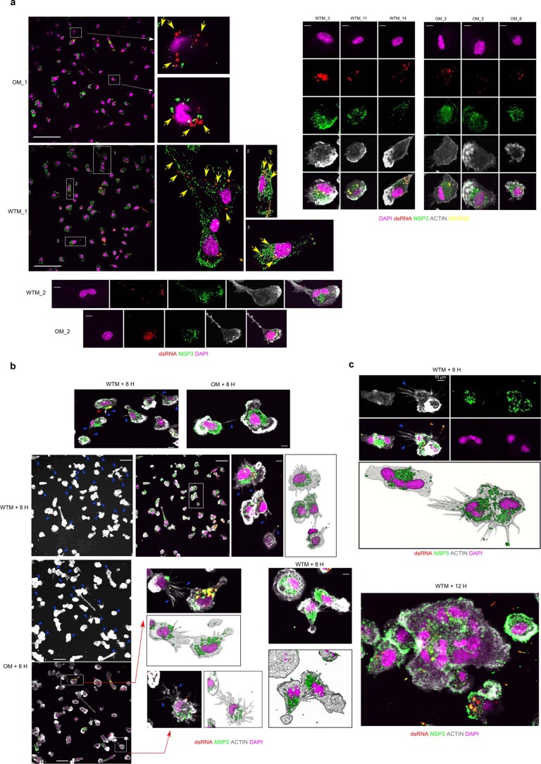
Extended Data Fig. 3. Viral compound detection in cultured BALF macrophages.
(a) Confocal microscopy images show the intracellular localization and accumulation of SARS-CoV-2 nonstructural protein 3 (NSP3) and double-stranded RNA (dsRNA) in BALF CD64+ Mac isolated from WTM and OM at ≥221 days p.i. After 8 hours of culture, cells were fixed with 4% formaldehyde. Immunostaining included DAPI (nucleus; purple), anti-dsRNA (red), and anti-NSP3 (green). Yellow arrows highlight dsRNA within the macrophages. (b) Similar to (a), these confocal microscopy images depict the intracellular localization of NSP3 and dsRNA in BALF CD64+ Macs, with the addition of Phalloidin (white) staining to highlight protrusions within the macrophages. Blue arrows indicate the presence of protrusions. Increased threshold on fields displaying only Actin was used to facilitate visualization of pseudopod- and TNT-like structures. (c) Confocal microscopy images as in (b) show syncytia observed in MAC culture. Scale bars in (a) = 45 µm, Scale bars in (b) and (c) = 35 µm. Some images use 3D rendering techniques to highlight the three-dimensional aspect of the visualization.

