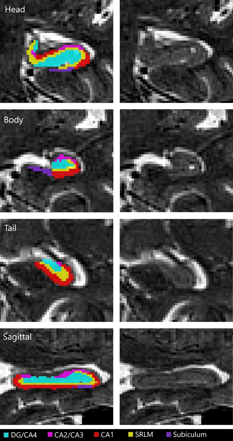
Figure 1.
An example of hippocampal segmentation resulting from the MAGeT-Brain algorithm over a T2-weighted MRI from a TRIAD subject. A sagittal slice and coronal slices of the hippocampus head, body and tail are displayed with and without a subfield mask overlay. CA, cornu ammonis; DG, dentate gyrus; SRLM, strata radiatum, lacunosum and moleculare.

