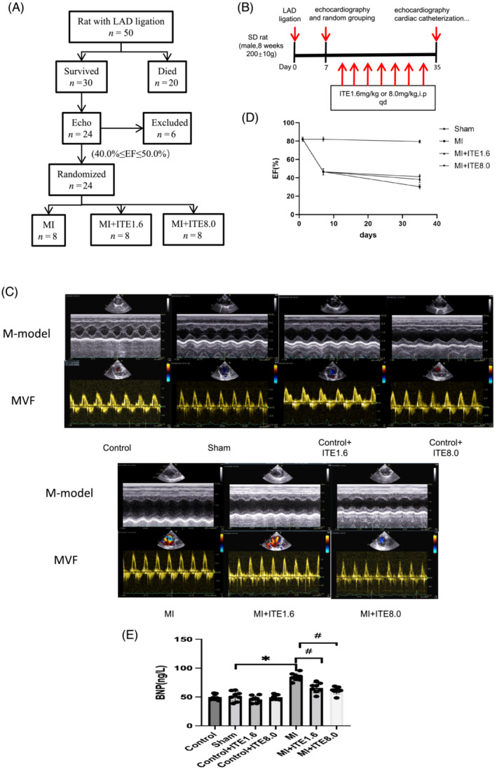
Figure 1.

ITE ameliorates cardiac dysfunction and heart failure in MI rats. (A) Flow chart of inclusion criteria based on echocardiographic ejection fraction (EF) used to obtain a cohort of MI mice with similar levels of cardiac dysfunction that were then randomized to receive vehicle or ITE. (B) Schematic diagram describing the animal experiments. (C) Representative M‐mode echocardiograms and mitral valve inflow (MVF) from control, sham, control + ITE1.6, control + ITE8.0, MI, MI + ITE1.6, and MI + ITE8.0. (D) The EF timeline shows the changes in EF at different time points in sham, MI, MI + ITE1.6, and MI + ITE8.0 rats. (E) Serum BNP levels measured by ELISA. N = 6–8 per group. Data are presented as mean ± SD. *P < 0.05 vs. sham group. # P < 0.05 vs. MI group.
