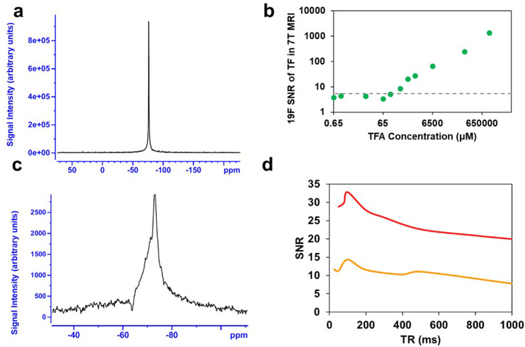
Figure 5.
(a). Representative in vitro TFA spectra (100%, 13 mM) were acquired using our experimental setup and custom RF coil on a 7-T animal MRI scanner (300 MHz) employing a single pulse sequence. (b) Corresponding 19F SNRs at 7-T MRI are presented for serial dilutions of 100% TFA (green), progressively diluted until reaching the limit of detection (LOD) threshold indicated by dashed lines. (c) Representative 19F MR scan (scan time = 4 min, TR = 80 ms). (d) SNR of Q2TFL obtained from 19F MRS using a 7-T MRI scanner, with scans acquired under both 4 min (red) and 1 min (orange) scan times, while varying the TR.

