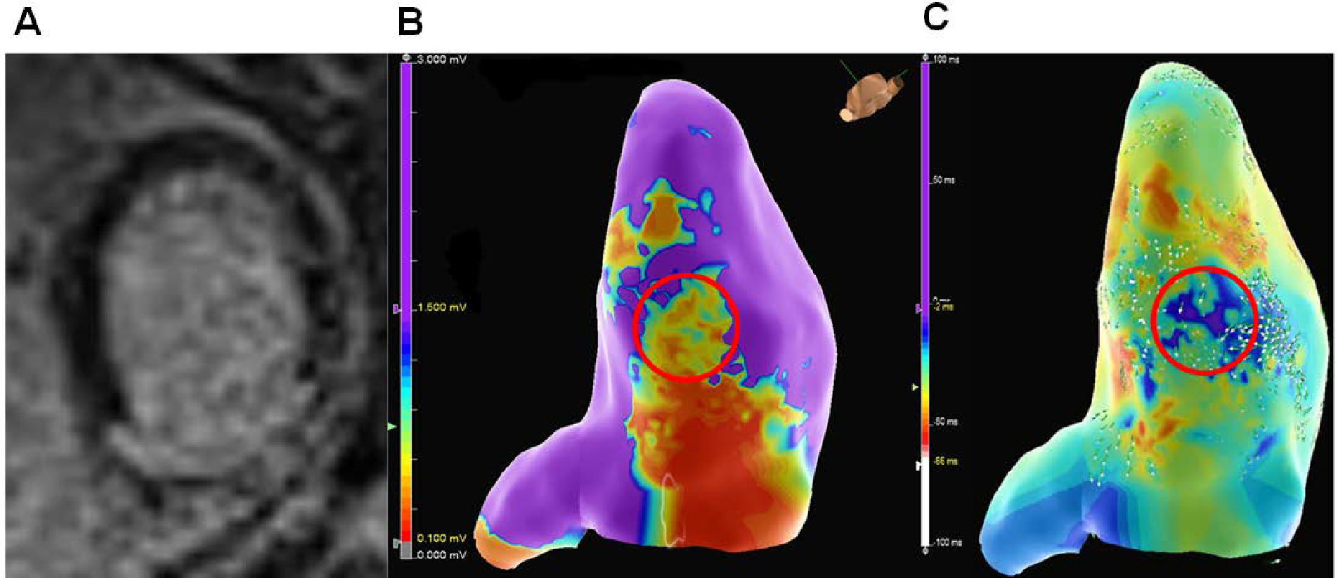
Figure 2:

A patient with an ICM presenting with ES secondary to inferior MI, resulting in inferior left ventricular scar as shown by (A) late gadolinium enhancement on cardiovascular magnetic resonance imaging. (B) The imaging correlated with areas of low voltage (red circle) on electroanatomical mapping during VT ablation in the inferior left ventricle. (C) Activation mapping in sinus rhythm demonstrated areas of late activation and slow conduction (red circle) that corresponded to the area of scar.
