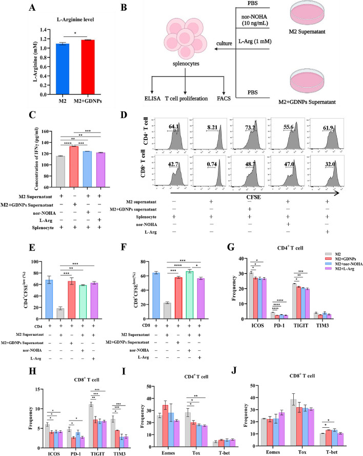
Fig. 3.
GDNPs reduced ARG1 production to promote proliferation and activation of T cells by polarizing TAMs. a L-Arginine level in supernatants from M2 and M2 + GDNPs was detected by L-Arginine Assay Kit. b Schematic diagram of culture system. c IFN-γ concentration in the supernatants was analysed by ELISA. d ARG1 inhibited the proliferation of CD4 + and CD8 + T cells. Splenocytes were cultured with M2 supernatant supplemented with PBS or nor-NOHA (10 ng/mL) or L-Arginine (1 mM) and M2 + GDNPs supernatant for 72 h. Representative histograms of CD4 + and CD8 + T cells proliferation. e, f Quantification of CD4 + and CD8 + T cells proliferation using CFSE dilution. g, h, i, j Splenocytes were cultured with M2 supernatant supplemented with PBS or nor-NOHA (10 ng/mL) or L-Arginine (1 mM) and M2 + GDNPs supernatant for 48 h. g, h Representative quantification of immune checkpoints of ICOS, PD-1, TIGIT, and TIM3 on CD4 + and CD8 + T cells. (i, j) The expression of transcription factors Eomes, Tox and T-bet in CD4 + and CD8 + T cells were analyzed. The results represent three independent experiments as the mean ± SEM. One-way ANOVA (c, g, h, i, j) and Student’s t test (a, e, f) were used to compare results of different experimental groups for statistically significant difference (*P < 0.05, **P < 0.01, ***P < 0.001, ****P < 0.0001)

