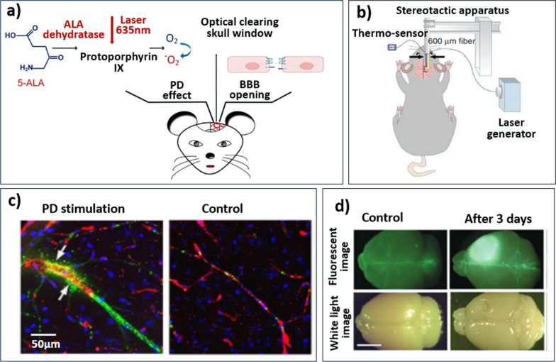
Fig. 2.
a Schematic of photodynamic therapy-induced BBB opening in an animal model, adapted from Zhang et al. [68]. 5-ALA, a photosensitising agent, is injected intravenously prior to light exposure. Through a small opening/window in the skull, light at an agent-specific wavelength (here 635 nm) is emitted into the brain leading to photochemical reactions and heating of the stimulated area. The thermal effect leads to the local opening of the BBB post-exposure. b Schematic depiction of the LITT delivery system in mice. The laser fibre (right arrow) is positioned 1 mm caudal to the thermo-sensor (left arrow). In contrast to PD, a photosensitising agent is not used and laser treatment is delivered via laser fibre, avoiding a skull window. Figure adapted from Salehi et al. [87] c experimental results of PD stimulation: confocal imaging of GM1-liposomes the with usage of markers of the neurovascular unit. Cell nuclei are labelled with DAPI, liposome leakage outside the vascular endothelial cells are labelled by antibodies. The white arrows show the sites of liposome leakage. Images were taken from Zhang et al. [68]. d LITT increases BBB and BTB permeability in vivo. Representative white light and fluorescence images of mouse brains harvested on the indicated days after intravenous fluorescein injection. LITT was performed in the right somatosensory cortex. Control = unmanipulated brain. Scale bar = 5 mm, taken from Salehi et. al. [87]

