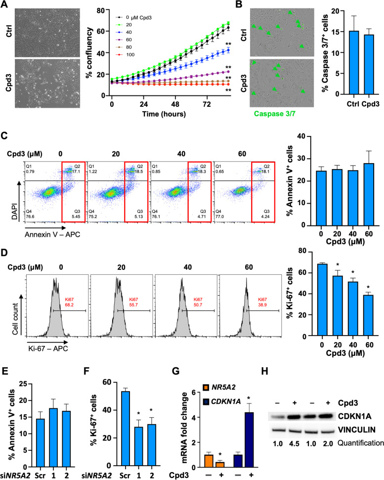
Fig. 2.
NR5A2 controls proliferation in differentiated cancer cells and is drugable. A Cell density and morphology were assessed after 72 h of treatment with Cpd3 (40 µM) versus control (Ctrl). Representative results for PDX215 cultures are shown (left). Additionally, the overall cell confluency was monitored over an 80-h period using the IncuCyte® platform (right). B Caspase 3/7 staining for cells treated with control or Cpd3 (40 µM) for 72 h. Quantification and representative images for PDX215 from n = 5 experiments are presented. C Apoptosis analysis was performed using DAPI/Annexin V flow cytometry in PDX215 cells treated with graded doses of Cpd3. The lower right quadrant indicates early apoptosis, while the upper right quadrant represents late apoptosis (marked by the red rectangle). Quantification reveals the percentage of combined early and late apoptotic cells (n = 3). D Flow cytometric dot blot analyses were carried out to examine Ki-67 expression after 72 h of treatment with graded doses of Cpd3 in PDX215 cells. The quantification depicts the percentage of Ki-67+ cells (n = 3). E Apoptosis analysis, measured by DAPI/Annexin V flow cytometry, and the measurement of proliferation (F), indicated by the number of Ki67+ cells, were conducted in PDX215 cells treated with scramble siRNA and the two most effective siNR5A2 variants (#1 and #2) for 72 h. Quantification of four biological replicates is displayed. G qPCR fold change of NR5A2 and CKDN1A (p21) mRNA following 72-h treatment with Cpd3 (80 µM) in PDX215 cells. H Western blot analysis of CDKN1A protein levels following 72 h treatment with Cpd3 (80 µM) in PDX215 cells. Data were presented as mean ± SD and statistically analyzed using two-tailed Mann–Whitney tests. Asterisks indicate significance at the indicated levels: * p < 0.05 and ** p < 0.01. Please also see Supplementary Fig. 2

