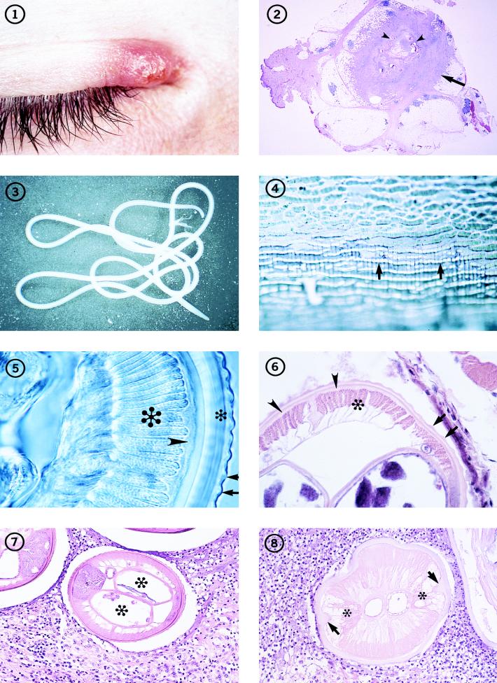
FIG. 1-8.
Individual with an inflammatory lesion at the inner canthus of the eye from which an immature adult D. tenuis worm was removed.
Fig. 2 Subcutaneous nodule (arrow) removed from a patient in Florida, containing several sections of a D. tenuis worm (arrowheads). Hematoxylin and eosin stain; magnification, ×4.5.
Fig. 3 An immature adult female Dirofilaria species that measured more than 12 cm in length, removed intact from human subcutaneous tissues.
Fig. 4 The surface of the cuticle of D. repens, illustrating the beaded appearance of the longitudinal ridges (arrows). The worm was preserved and mounted in glycerin. Magnification, ×182.
Fig. 5 Section through an adult D. repens worm, illustrating the appearance of the longitudinal ridges (arrows) in transverse section, as well as the thick, multilayered cuticle (small asterisk), hypodermis (arrowhead), and muscle cells (large asterisk). The worm is unstained but cleared in glycerin. Magnification, ×391.
Fig. 6 Portion of a transverse section of D. tenuis, showing the longitudinal ridges (arrows) on the surface of the multilayered cuticle, the thin hypodermis (arrowheads), and the coelomyarian muscles (asterisk). Hematoxylin and eosin stain; magnification, ×227.
Fig. 7 Transverse section through a female D. tenuis worm removed from human subcutaneous tissues, illustrating its characteristic morphologic features. The paired uterine tubes (asterisks) contain unsegmented eggs. Hematoxylin and eosin stain; magnification, ×121.
Fig. 8 Dead, degenerating female Dirofilaria worm, with a thickened cuticle, prominent lateral internal cuticular ridges (arrows), and frothy, vacuolated lateral chords (asterisks). In the pseudocelom, the tube to the left is the vagina; adjacent to it is the intestine. This section is cut in the anterior one-fifth of the worm. Hematoxylin and eosin stain; magnification, ×164.

