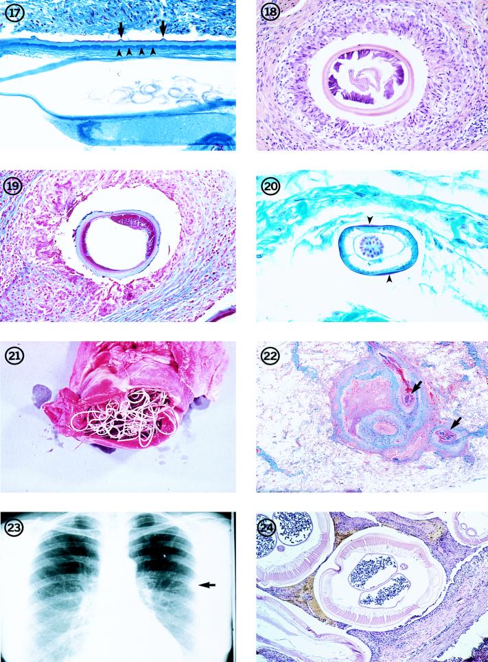
FIG. 17-24.
Longitudinal section through a female O. cervicalis worm from a naturally infected horse, illustrating the morphologic features of the cuticle, including the outer circular ridges (arrows) and the inner striae (arrowheads). There are four striae per ridge. Trichrome stain; magnification, ×209.
Fig. 18 Transverse section through a female Onchocerca worm removed from a human in Japan. The variable thickness of the cuticle is evident, as is the weak musculature. Two uterine reproductive tubes are present in the body cavity. Hematoxylin and eosin stain; magnification, ×114. (From the case presented in reference 22.)
Fig. 19 Transverse section of the female Onchocerca worm illustrated in Fig. 16. Note the unevenly thick cuticle and the low, weak, poorly developed musculature. Trichrome stain; magnification, ×114. (From the case reported in reference 17.)
Figure 20 Section through a male O. gutturosa worm from a naturally infected cow, illustrating the thin cuticle which thickens laterally (arrowheads), small number of muscle cells, and large testis, which fills the body cavity. The intestine is the small, collapsed tube adjacent to the testis. Trichrome stain; magnification, ×328.
Fig. 21 Adult D. immitis in the right ventricle of the heart of its natural host, the domestic dog. Reprinted from reference 62 with permission of the publisher.
Fig. 22A coin lesion in a human lung, involving a small pulmonary artery. Sections of an immature D. immitis worm (arrows) can be seen in the obstructed vessel. Hematoxylin and eosin stain; magnification, ×3.6.
Fig. 23 Chest X ray showing a typical coin lesion (arrow) in a human lung as a result of a D. immitis infection.
Fig. 24 Transverse section of an adult female D. immitis worm, illustrating the characteristic morphologic features of this parasite. Hematoxylin and eosin stain; magnification, ×45.

