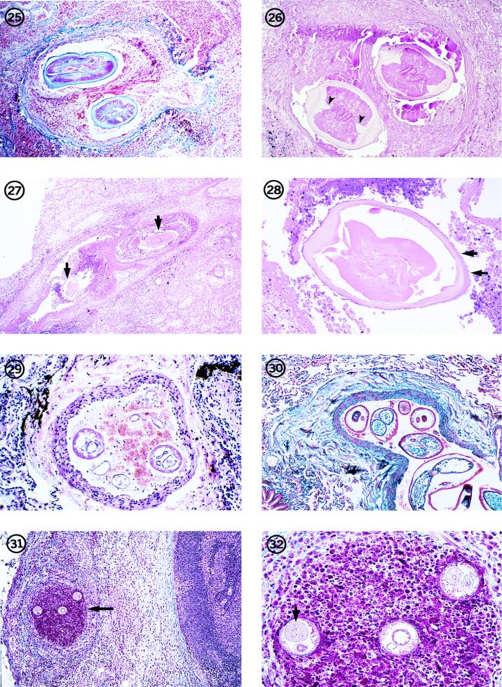
FIG. 25-32.
Dead D. immitis worm in an occluded small pulmonary artery in a human lung. The parasite can be identified by its morphologic features, including its size and the thick but smooth cuticle with prominent lateral, internal cuticular ridges. Hematoxylin and eosin stain; magnification, ×45.
Fig. 26 Dead, degenerating, female D. immitis worm in a small pulmonary nodule. Note the thick, swollen cuticle with prominent internal cuticular ridges (arrowheads). The three tubes in the pseudocelom (two reproductive tubes and an intestine) indicate that it is a female worm. Hematoxylin and eosin stain; magnification, ×91.
Fig. 27 Low-power view of sections of D. repens (arrows) in an obstructed pulmonary artery of an individual in Italy. Hematoxylin and eosin stain; magnification, ×11.4. Reprinted from reference 62 with permission of the publisher.
Fig. 28 Higher magnification of one of the sections of the worm in Fig. 27. The parasite is in an advanced state of degeneration as indicated by the swollen cuticle and degeneration of the soft tissues of the worm. However, longitudinal ridges can be seen on the cortical surface of the cuticle (arrows). Hematoxylin and eosin stain; magnification, ×91. Reprinted from reference 62 with permission of the publisher.
Fig. 29 Section through a small pulmonary artery removed from an individual who had lived in India, showing two transverse sections through a Brugia-like worm. Hematoxylin and eosin stain; magnification, ×91. Reprinted from reference 62 with permission of the publisher.
Fig. 30 W. bancrofti-like worm lying in a small pulmonary artery of a soldier who had served in Singapore. Trichrome stain; magnification, ×45. (From the case presented in reference 15.)
Fig. 31 Section through a peripheral lymph node from the upper arm of a patient in Ohio. Three sections of a small Brugia worm are present in an occluded lymph channel (arrow) lying inside the nodular capsule. The medullary portion of the lymph node is visible to the right of the nodule. Hematoxylin and eosin stain; magnification, ×59.
Fig. 32 Higher magnification of the worm in Fig. 31. Two sections through the anterior end at the level of the esophagus and ovejector (right and left sections) and one section through the region of the intestine and vagina uterina (middle section) are evident. The thin body wall, triradiate esophagus (arrow), few muscle cells per quadrant, and the muscular vagina vera and uterina are evident. Hematoxylin and eosin stain; magnification, ×237.

