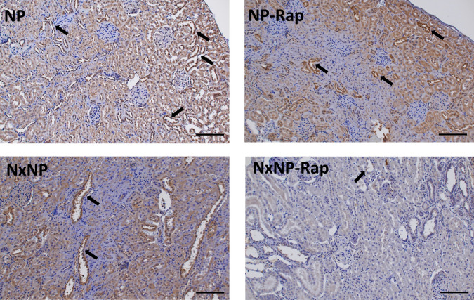
Fig 7. α-Klotho Renal immunohistochemistry.
Immunohistochemistry for ⍺-klotho, in renal tissue from rats fed 0.6% phosphorus with intact (NP) and reduced (NxNP) renal function receiving either placebo or rapamycin (Rap). Positive staining (brown color) was located in the cytoplasm of tubular cells of the kidney cortex, with preferential expression in distal tubules (arrows). Scale bar = 100 μm.

