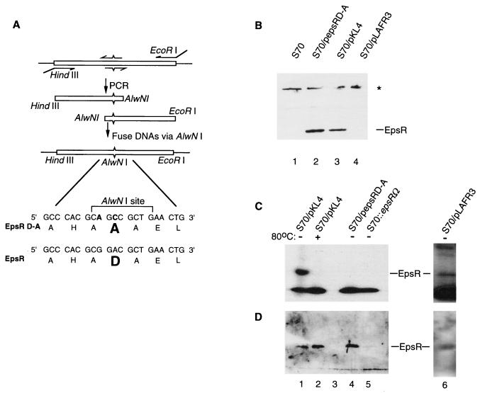
FIG. 2.
(A) A schematic diagram of site-directed mutagenesis of the conserved aspartate (D47) in epsR is shown. Oligonucleotide primers are indicated with arrows. The internal pair of primers contains changes in the epsR sequence, indicated by ^ or ˇ. Changes made in the epsR sequence are shown at the bottom of the figure, and the encoded amino acids are in the standard one-letter code. The original aspartate in the EpsR sequence and the alanine in the EpsRD-A sequence are shown in bold type. (B) Western blot analysis shows that EpsRD-A is expressed in R. solanacearum (S70) when cells are transformed with pepsRD-A. The star designates nonspecific recognition of protein by the anti-EpsR antibodies. (C) EpsR (lane 1), but not EpsRD-A (lane 3), is phosphorylated. A longer exposure of this gel (lane 6) reveals the phosphorylation of chromosomally derived EpsR. (D) After autoradiography, the gel in Fig. 3C was subjected to Western blot analysis with anti-EpsR serum (21). The results show that EpsR and EpsRD-A are present in cell lysates in similar quantities.

