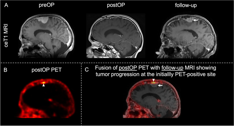
Fig. 2.
Case example of tumor progression. MRI and PET imaging in patients with tumor progression on PET. A Preoperative and postoperative sagittal contrast-enhancing T1-weighted MRI demonstrating homogenous contrast enhancement of a left hemispheric convexity meningioma with no apparent residual tumor after resection. Follow-up MRI showing consequent tumor recurrence at the same site (arrow). B Sagittal postoperative [68Ga]Ga-DOTA-TATE PET/CT scan of the same patient demonstrating residual tumor after resection (arrowhead). C Fusion images of postoperative PET scans and latest contrast-enhancing T1-weighted follow-up MRI showing a correlation between residual tumor on PET (arrowhead) and tumor progression on MRI (arrow). Abbreviations: ceT1 contrast-enhancing T1-weighted MRI; preOP preoperative; postOP postoperative

