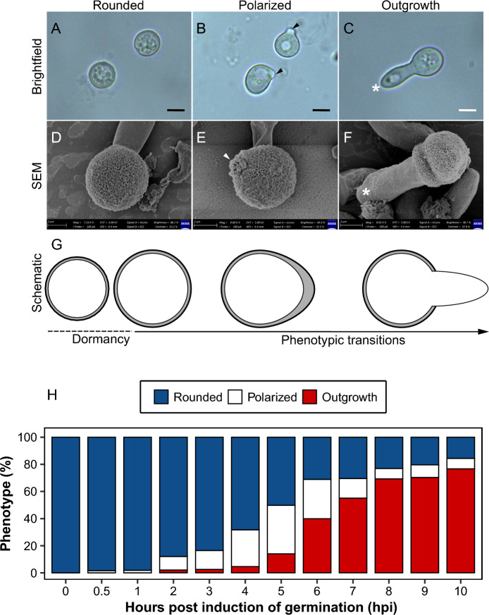
Fig. 1.
Bright-field and scanning electron microscopy micrographs showing the transitions during germination of chlamydospores of Fusarium oxysporum f. sp. cubense Tropical Race 4 (Foc TR4): rounded (A, D), polarized growth (B, E), and outgrowth (C, F). The stages of germination are further illustrated in the schematic (G). The site of polarity establishment (indicated by arrow heads) on polarized chlamydospores, is the point where the germ tube (indicated by white asterisk) emerges during outgrowth. Scale bar = 20 µm. Phenotypic changes during the process of germination of chlamydospores in Foc TR4 (H)

