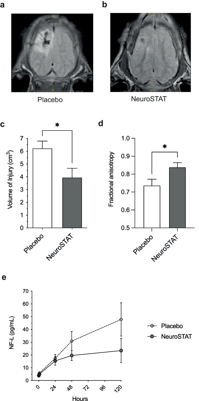
Fig. 1.
Translational efficacy outcomes of NeuroSTAT in a piglet study with controlled cortical impact injury. Neuroimaging on day 5 post-injury depicting A magnetic resonance imaging anatomical images representative of the median injury in the NeuroSTAT-treated group and B in the placebo group. C Volume of injury measured by manual tracing on each slice of area of increased signal abnormality on FLAIR imaging by board-certified neuroradiologist blinded to treatment group. D Fractional anisotropy in peri-contusional tissue using diffusion tensor imaging. E Neurofilament light (NF-L) in serum day 1–5 post-injury. Data are presented as mean and SEM. *p<0.05. Adapted from [29, 30] with permission from the publisher

