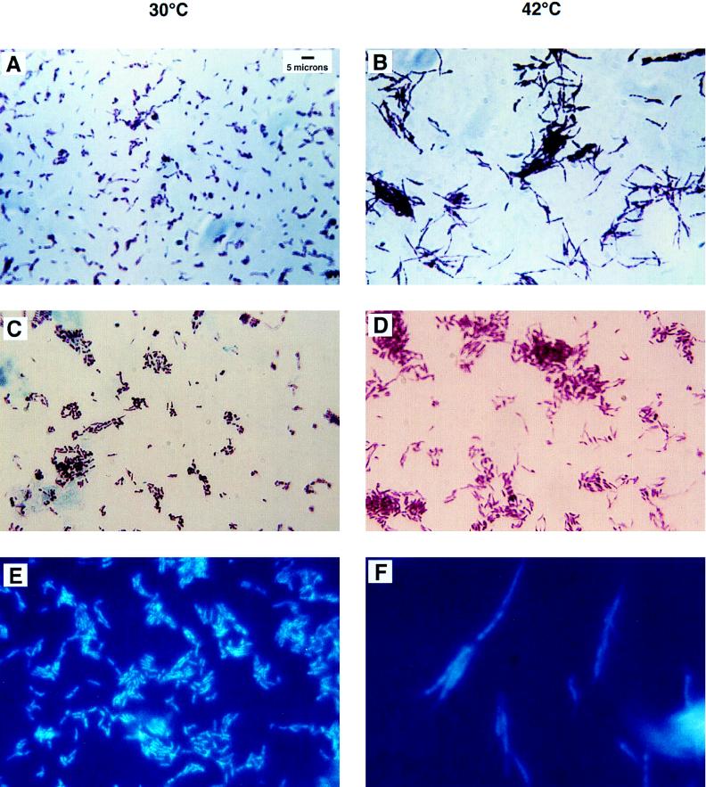
FIG. 1.
Acid-fast stains of SMEG 1 after growth at the permissive and nonpermissive temperatures for 6 h. (A) SMEG 1 cells incubated at 30°C appear as normal bacilli which average 2 to 3 μm in length. (B) SMEG 1 cells appear long and filamentous after 6 h of incubation at 42°C. (C) SMEG 1(pAK30) at 30°C shows the wild-type phenotype with additional copies of dnaG provided on an extrachromosomal plasmid. (D) SMEG 1(pAK30) at 42°C exhibits a wild-type phenotype showing that it has been rescued by the wild-type copy of dnaG provided by the plasmid pAK30. (E) SMEG 1 cells were incubated at 30°C and then stained with DAPI. These stained cells show nucleoids that are stained uniformly and are condensed and centrally located. (F) SMEG 1 cells after 6 h of incubation at 42°C have faintly stained, poorly defined, diffuse nucleiods.

