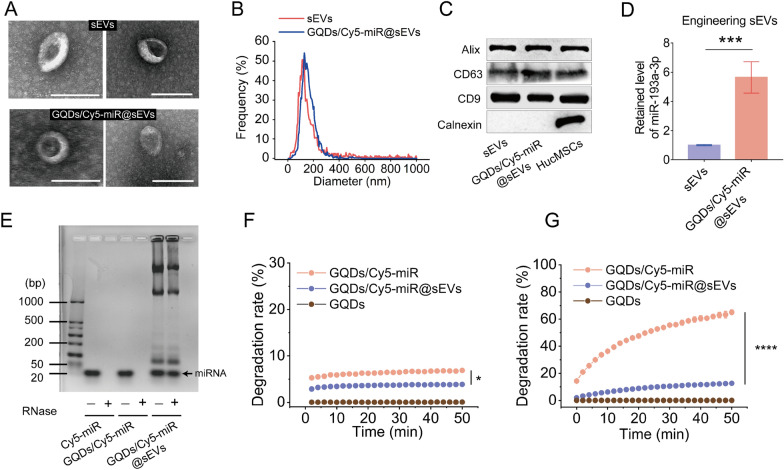
Fig. 3.
Characterization and drug loading capacity of GQDs/Cy5-miR@sEVs. A TEM images of sEVs (upper panel) and GQDs/Cy5-miR@sEVs (lower panel). Scale bar = 200 nm. B Size distribution of sEVs and GQDs/Cy5-miR@sEVs. C Expression of Alix, CD63, CD9, Calnexin in sEVs, GQDs/Cy5-miR@sEVs and HucMSCs was detected by Western blot. D MiR-193a-3p in GQDs/Cy5-miR@sEVs and sEVs were detected by qRT-PCR. E The degradation of Cy5-miR, GQDs/Cy5-miR and GQDs/Cy5-miR@sEVs in RNase solutions was detected by agarose gel electrophoresis. F Stability of GQDs/Cy5-miR and GQDs/Cy5-miR@sEVs in PBS in 50 min at room temperature. G Stability of GQDs/Cy5-miR and GQDs/Cy5-miR@sEVs in 50 min after being incubated within mouse serum at 37 °C. Data are expressed as mean ± SD and analyzed by one-way ANOVA. *p < 0.05, ***p < 0.001, ****p < 0.0001

