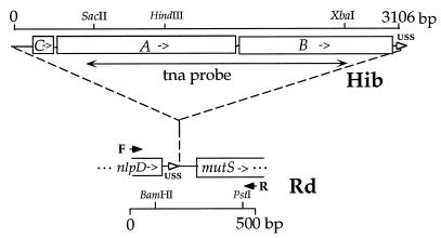
FIG. 1.
Map of tna insert region showing positions on Rd genome at which tna DNA is inserted and of unique restriction sites. The scale of the insert (Hib) map is 50% that of Rd. The primers used to amplify the insert region are depicted with solid arrows; F, nlpD-F; R, mutS-R. Open arrows depict paired USSs. The position of a SacII/XbaI fragment (bp 705 to 2680) used as a tna-specific probe is indicated.

