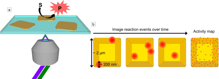
Figure 2.
Single-molecule fluorescence imaging of fluorogenic probes on nanoscale catalysts. (a) Schematic of objective-based TIRF microscopy in which a laser is sent through a TIRF microscope objective at an angle such that it is internally reflected by the glass coverslip. The catalyst converts an initially nonfluorescent substrate molecule (S) into a fluorescent product (P). The evanescent field from the TIRF illumination excites the fluorescent product, and photons emitted by the activated probe are collected by the objective. For semiconductor photocatalysts, dual excitation may be used in which one laser with a photon energy greater than the band gap of the semiconductor (e.g., 405 or 450 nm) excites electrons into the conduction band of the semiconductor, and a lower-energy laser (e.g., 488, 532, or 567 nm) excites the activated probe. (b) Schematic for superlocalization of activated probe molecules (red circles) on a faceted catalyst particle (shown in yellow). The emission profile for each fluorescent molecule is diffraction-limited, but the center position of the fluorophore can be localized with nanoscale precision given a sufficient number of photons are collected over the background and as long as two molecules within a diffraction-limited region are not emitting at the same time. By localizing the positions of many activated probes over time, super-resolution activity maps can be produced, which show how the activity varies at the nanoscale across the catalyst surface (right image in panel b).

