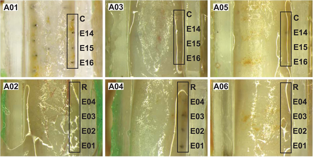
Figure 3.
End-of-study electrode assessment showing a top-down view into the silicone guide channel. Each image shows the gross position of electrodes at the outer edge of the sciatic nerve for each implanted animal subject (A01, A02, A03, A04, A05, A06). Images were taken after removal of the green silicone sealant covering the nerve, after fixation and removal of the sciatic nerve at post-implantation week 38.

