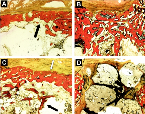
FIG. 5.
Histological sections demonstrating the healing of cortical bone. (A) Healed cortical window (black arrow) in negative control group; (B) extensive reactive new bone formation in cortical defect of untreated-infection group; (C) healed cortical window (white arrow) and associated intramedullary new bone formation (black arrow) in treated-infection group; (D) unhealed cortical defect with extruding biomaterial fillers in sham-treated group. Modified van Gieson stain. Magnification, ×25.

