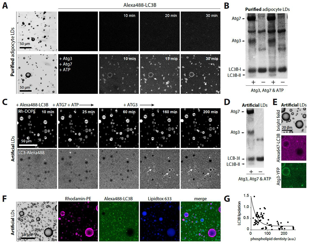
Figure 5. ATG3 lipidates LC3 to purified and artificial LDs.

A. Confocal imaging of purified adipocyte LDs in HKM buffer containing Alexa488-LC3B, in the presence or absence of the lipidation reaction components ATG7, ATG3, ATP.
B. LDs from the previous experiment are collected and analyzed using SDS–PAGE in a stained Coomassie blue.
C. Confocal imaging of triolein-in-buffer droplets decorated by PC/PE (7/3) incubated with Alexa488-LC3B, then ATG7 and ATP. No lipidation occurred. When ATG3 was subsequently added, lipidation occurred on the artificial LDs (arrows show examples).
D. Artificial LDs from the previous experiment are collected and analyzed using SDS–PAGE in a stained Coomassie blue.
E. Triolein-in-buffer droplets decorated with PC/PE at different monolayer phospholipid densities (based on Rho-PE signal) are imaged using confocal microscopy after being incubated with Alexa647-LC3B and Atg3/Atg3-YFP (80/20), ATP, and ATG7.
F. Confocal imaging of triolein-in-buffer droplets decorated by PC/PE (7/3) at different monolayer phospholipid densities varied from 0.005% to 0.2% (w/w to triolein). They are incubated with Alexa488-LC3B and Atg3 (80/20), ATP and ATG7.
G. Quantification of F. LC3B-Alexa488 lipidation to triolein droplets as a function of the phospholipid density.
See also Figure S5.
