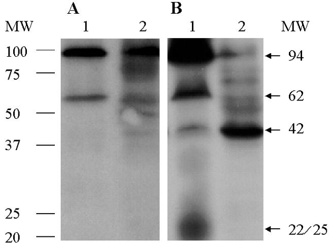
FIG. 1.

Results of chemiluminescence assays using whole-cell lysates (A) and membrane preparations (B) of human B. pilosicoli strain SP16 (lanes 1) and E. coli (lanes 2) reacted with ampicillin conjugated to digoxigenin followed by immunoblot and chemiluminescence assays. Two prominent PBPs with molecular masses of approximately 94 and 62 kDa and a minor 42-kDa reactive band present in B. pilosicoli migrate at a position similar to the high-molecular-weight PBP-1 and PBP-3 and the low-molecular-weight PBP-5/6 of E. coli. A less-defined band, of approximately 22 to 25 kDa, that is present in the membrane preparation of B. pilosicoli (panel B) but not in the intact cells (panel A), might represent a breakdown product of high-molecular-weight PBPs. MW, molecular weight in thousands.
