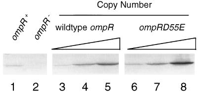
FIG. 2.
Western immunoblot analysis of OmpR levels in the cells. Cells were grown overnight in LB medium without (lanes 1 and 2) or with (lanes 3 to 8) ampicillin (50 μg/ml), subcultured into 40 ml of the same medium, and grown to mid-log phase. Total cellular protein was prepared as described previously (10). One hundred micrograms of total protein from each sample was separated by SDS–10% PAGE and then transferred to a polyvinylidene difluoride membrane. Western blot analysis was then performed with rabbit anti-wild-type OmpR antiserum (1:5,000) and alkaline phosphatase-conjugated goat anti-rabbit antibody (1:10,000). The immune complexes were detected with the Vistra ECF substrate (Amersham Life Science Inc.) and visualized with a FluorImager Storm 840 system (Molecular Dynamics). Lanes: 1, MC4100; 2, LM101; 3, pLAN701 in CYL303; 4, pLAN701 in LM101; 5, pLAN801 in LM101; 6, pLAN702 in CYL303; 7, pLAN702 in LM101; 8, pLAN 802 in LM101.

