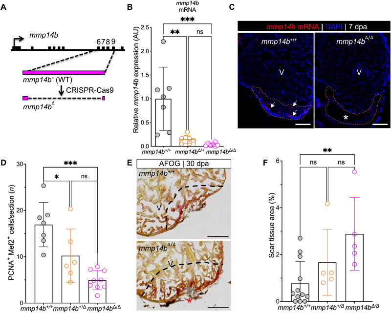
Fig. 2. Inactivation of the mmp14b gene results in reduced cardiomyocyte proliferation and impaired scar resolution in response to heart injury in zebrafish.
(A) Strategy used to create an mmp14b-null allele. (B) Relative mmp14b expression in mmp14b+/+, mmp14b+/Δ, and mmp14bΔ/Δ larvae as measured by qPCR. (C) RNAscope in situ hybridization for mmp14b mRNA in mmp14b+/+ and mmp14bΔ/Δ hearts at 7 dpa. Arrows, mmp14b expression in the wild-type mmp14b+/+ heart following injury. The asterisk denotes lack of evident mmp14b expression in the mmp14bΔ/Δ heart. (D) Cardiomyocyte proliferation indices at 7 dpa. (E) Representative frontal sections of mmp14b+/+ and mmp14bΔ/Δ hearts collected 30 dpa and stained with Acid Fushin Orange G (AFOG) to detect muscle (brown), fibrin (red), and collagen (blue). The asterisk highlights collagen-rich scar tissue. (F) Percent of ventricle composed of scar tissue at 30 dpa in mmp14b+/+, mmp14b+/Δ, and mmp14bΔ/Δ hearts. Data were collected for five to eight sections per heart and averaged to generate each data point. The black dashed lines mark the injured area. Scale bars, 100 μm. Statistical significance was determined using one-way analysis of variance (ANOVA) with Tukey’s multiple comparisons test. ns, not significant; *P < 0.05; **P < 0.01; ***P < 0.001.

