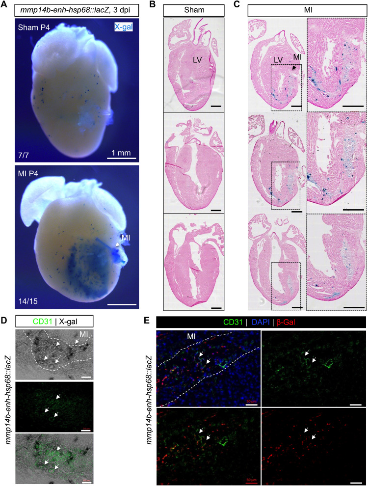
Fig. 6. Zebrafish mmp14b-enh is induced by myocardial injury in neonatal mice.
(A) Whole-mount and coronal sections (B and C) of representative mmp14b-enh-hsp68::lacZ hearts in control (sham) and injured myocardial infarction (MI) neonatal mice. Myocardial infarction was induced by permanent coronary ligation in neonatal mice on P1, and β-galactosidase (β-gal) expression was assessed by X-gal staining on P4. Sections were counterstained with hematoxylin and eosin. Note the activation of β-galactosidase (blue staining) in injured mmp14b-enh-hsp68::lacZ hearts (14 of 15) but not in sham-operated mmp14b-enh-hsp68::lacZ (7 of 7). Boxes in [(C), left] are shown at higher magnification in [(C), right]. LV, left ventricle. (D and E) Coronal sections of mmp14b-enh-hsp68::lacZ neonatal mouse hearts stained for X-gal (gray) (D) or immunostained for β-galactosidase (red) (E) and immunostained for the endothelial cell marker CD31 (green) at 3 dpi. Colocalization of X-gal and CD31 is shown in the bottom of (D). Colocalized expression is highlighted by white arrows. The infarcted area is outlined with a white dashed line. DAPI staining (blue) is shown in (E). Scale bars, 500 μm [(B) and (C)] and 50 μm [(D) and (E)].

