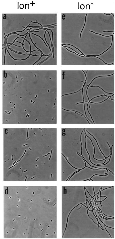
FIG. 3.
Phenotypes of minC mutants in the presence of MinD in lon+ and lon mutant cells. Representative examples are shown; other results are summarized in Table 1. The lon+ ΔminCDE hosts were PB114 (λDB164 Plac-minD) (a to c) and RC3F−/pDB211 (PaadA-minD) (d). The lon ΔminCDE hosts were MS114 (λDB164) (e to g) and MS114/pDB211 (h). The strains contained minC plasmid pJPB120m4 (PaadA-minC24) (a and e), pJPB120m11 (PaadA-minC27) (b and f), pMS709 (Plac-minC28) (c and g), or pJPB120m5 (PaadA-minC25) (d and h). Cells were grown overnight in the presence of 0.2 mM IPTG (isopropyl-β-d-thiogalactopyranoside) prior to examination by phase-contrast microscopy (2).

