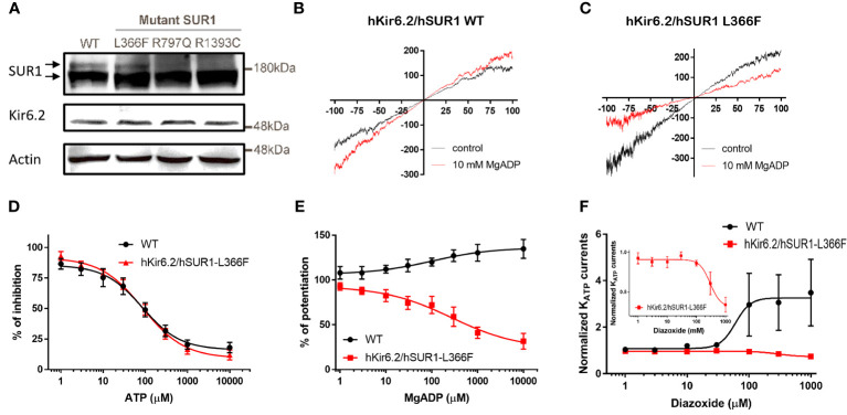
Figure 1.
(A) Western blots show total SUR1 levels in HEK293 cells. SUR1 signals exhibit an upper band that corresponds to a fully glycosylated KATP channel (upper arrow), while the lower band represents immature KATP channels that are still stuck intracellularly (lower arrow) in wild-type (WT) SUR1. The upper bands are visible in HEK293 cells transfected with WT hSUR1 and hSUR1-L366F but absent in HEK293 cells transfected with hSUR1-R797Q and hSUR1-R1393C. β-actin serves as the loading control. (B, C) Representative voltage-clamp recording traces from inside-out patches containing KATP channels. Adding 10 mM MgADP (red traces) to the intracellular side potentiated WT hsSUR1 (B) but inhibited hSUR1-L366F (C) containing KATP currents. (D) WT and hSUR1-L366F containing KATP channels exhibited comparable sensitivity to the intracellular ATP (WT: IC50 = 85.4 µM, Hill slope = 0.97, n = 4; hSUR1-L366F: IC50 = 86.1 µM, Hill slope = 0.87, n = 6–8). (E) Intracellular MgADP potentiated WT KATP channels but inhibited hSUR1-L366F KATP (WT: EC50 = 102 µM, Hill slope = 0.65, n = 4–5; hSUR1-L366F: IC50 = 268 µM, Hill slope = 0.59, n = 3). (F) Diazoxide potentiates WT KATP Currents (EC50 = 60.1 μM, n = 5) but inhibits hSUR1-L366F KATP currents (IC50 = 297.7 μM, n = 4). The insert displays an enlarged dose-response curve of diazoxide on hSUR1-L366F KATP currents to highlight the inhibitory effect of diazoxide.

