
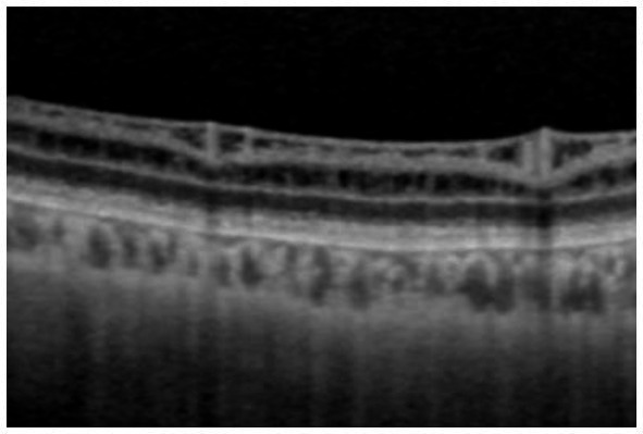
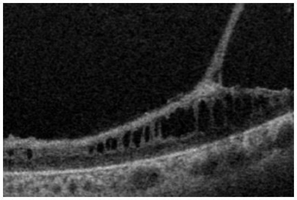
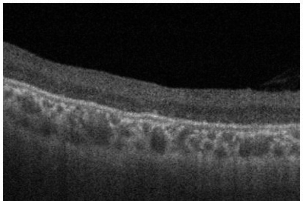
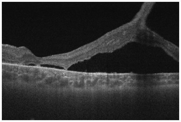
Table 2.
Grading of the peripheral fundi in patients with congenital X-linked retinoschisis.
| Grade | Notes | Ophthalmoscopy in the periphery | OCT in the periphery | Representative OCT images |
|---|---|---|---|---|
| Grade 1 | No retinoschisis | Normal with/without silver-gray reflex | No schisis, no cystoid changes, and normal layer structure |
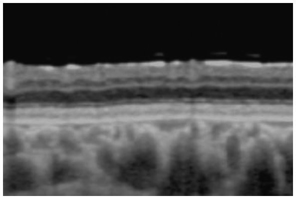
|
| Grade 2 | Occult retinoschisis | Normal with/without silver-gray reflex | Minor schisis and cystoid changes in GCL/INL |
|
| Grade 3 | Retinoschisis with retinal degeneration | Retinoschisis, shallow or bullous | Splitting in GCL and cystoid changes in INL |
|
| Grade 4 | Retinal atrophy, most likely atrophic outer leaf of the retina | Retinal degeneration | Atrophy of the retina with no layer structure |
|
| Grade +D | Retinal detachment with/without retinoschisis | Retinal detachment with/without retinoschisis | Retinal detachment with/without splitting/cystoid changes in GCL/INL |
|