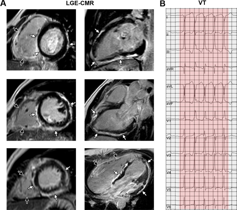
Fig. 2.
Ringlike LGE and corresponding electrocardiograph. A 25-year-old man had palpitation for 4 years with genetically diagnosed Bi-ACM (DSP mutation). A LGE-CMR images showed the extensive subepicardial fibrosis with a ringlike pattern involving the LV free wall and septum in both the short axis (left panel: from base to apex) and the long axis view (right panel: from 2 chamber to 4 chamber) (white arrowheads). Meanwhile, extensive LGE was also seen in the right ventricular wall (black arrowheads). B The 12-lead electrocardiograph showed monomorphic VT. LGE late gadolinium enhancement, Bi-ACM biventricular arrhythmogenic cardiomyopathy, DSP desmoplakin, CMR cardiac magnetic resonance, LV left ventricular, VT ventricular tachycardia

